 1 / 2
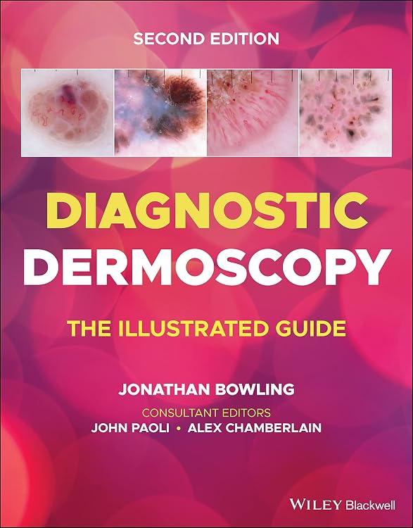
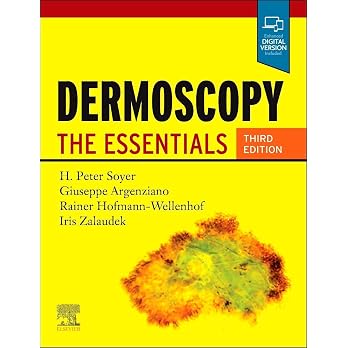
Diagnostic Dermoscopy: The Illustrated Guide, 2nd Edition
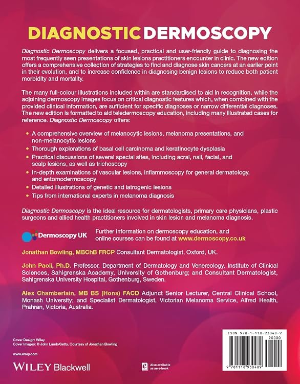
Diagnostic Dermoscopy: The Illustrated Guide is a second-edition clinical reference focused on recognising and diagnosing skin lesions with dermoscopy. Its full-colour clinical and dermoscopic images, case examples, and coverage of varied lesion types make it relevant to practitioners seeking a visual learning resource.
At a glance
- Combines standardised full-colour clinical illustrations with dermoscopy images focused on diagnostic features.
- Covers melanocytic and non-melanocytic lesions, melanoma, basal cell carcinoma and keratinocyte dysplasia.
- Includes specialised topics such as acral, nail, facial and scalp lesions, trichoscopy, vascular lesions and inflammoscopy.
- Described as formatted to support teledermoscopy education and illustrated case reference.
Reference price: $98.95
Price checked: Nov 19, 2026
View offerAhead of Black FridayExplore the selectionOften bought together
Pros and cons
Pros
- Broad topic range gives readers material on both common presentations and specialised anatomical sites.
- Image-led cases pair clinical information with dermoscopic features, supporting visual reference and study.
Technical specifications
9 specifications| Publisher | Wiley-Blackwell |
|---|---|
| Edition | 2nd |
| Publication date | July 12 2022 |
| Language | English |
| Print length | 336 pages |
| ISBN-10 | 1118930487 |
| ISBN-13 | 978-1118930489 |
| Dimensions | 21.34 x 1.27 x 27.18 cm |
|---|---|
| Weight listed | 215 g |
What to know before buying
- The publisher description presents this as a diagnostic and educational reference, not a substitute for clinical judgement or training.
Who is it for?
A good fit for
- Dermatologists and primary care physicians seeking a visual dermoscopy reference.
- Plastic surgeons and allied health practitioners involved in skin lesion or melanoma diagnosis.
- Learners who want illustrated cases spanning melanocytic, non-melanocytic and specialised-site lesions.
Less suited to
- Readers looking for a general dermatology textbook rather than a focused dermoscopy guide.
- Buyers who need a hands-on dermoscope or a digital course, neither of which is established as part of this book.
Customer reviews on Amazon
- 5 stars
82% 82% - 4 stars
14% 14% - 3 stars
2% 2% - 2 stars
0% 0% - 1 star
2% 2%
Aggregate ratings skew strongly positive, with most ratings in the five-star category, alongside a small one-star share. These figures offer a generally favourable but limited signal about reader reception, not evidence of specific praised features or explanations for dissatisfaction. The listing’s image-led format and broad diagnostic subject matter may suit the stated clinical audience, but ratings alone cannot confirm how useful the cases are in practice.
Frequently asked questions
What topics does the guide cover?
The listing describes coverage of melanocytic and non-melanocytic lesions, melanoma, basal cell carcinoma, keratinocyte dysplasia, vascular lesions and specialised sites including nails, face, scalp and acral areas.
Who is this book intended for?
The publisher identifies dermatologists, primary care physicians, plastic surgeons and allied health practitioners involved in skin lesion and melanoma diagnosis.
Does the guide include visual examples?
Yes. The description says it includes full-colour clinical illustrations, accompanying dermoscopy images and illustrated cases.
What edition and length are listed?
The listing identifies the book as the 2nd edition with a print length of 336 pages.
Alternative options
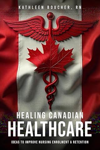
Healing Canadian Healthcare
$15.25
Anatomy & Physiology: The Best and Most Effective Way to Learn the Anatomy and Physiology of the Human Body: Workbook
$26.06
Fluids and Electrolytes: A Thorough Guide covering Fluids, Electrolytes and Acid-Base Balance of the Human Body
$24.99

The Eczema Relief Diet & Cookbook: Short-Term Meal Plans to Identify Triggers and Soothe Flare-Ups
$29.99
Medical Terminology: The Best and Most Effective Way to Memorize, Pronounce and Understand Medical Terms: Second Edition
$25.99
EKG/ECG Interpretation: Everything you Need to Know about the 12 - Lead ECG/EKG Interpretation and How to Diagnose and Treat Arrhythmias
$26.99

The Gambling Addiction Journal: A 90-Day Recovery Guide
$13.99
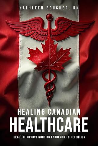
Healing Canadian Healthcare: Ideas to Improve Nursing Enrolment & Retention
$5.49
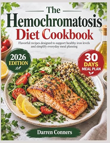
The Hemochromatosis Diet Cookbook: Flavorful recipes designed to support healthy iron levels and simplify everyday meal planning
$42.75
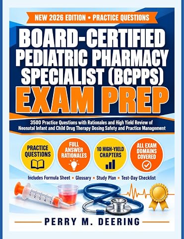
Board-Certified Pediatric Pharmacy Specialist (BCPPS) Exam Prep: 3500 Practice Questions with Rationales and High Yield Review of Neonatal Infant and Child Drug Therapy Dosing Safety and Practice Management
$48.94